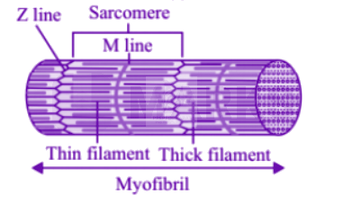
During muscle contraction in humans, the A band remains of the same size. Increase in Ca++level into the sarcoplasm leads to the binding of calcium with a subunit of troponin on actin filaments and thereby remove the masking on active sites for myosin. Utilising the energy from ATP hydrolysis, the myosin head now binds to the exposed active sites on actin to form a cross bridge. This pulls the attached actin filaments towards the centre of ' A ' band. The ' Z ' line attached to these actins are also pulled inwards thereby causing a shortening of the sarcomere, i.e., contraction. It is clear from the above steps, that during shortening of the muscle, i.e., contraction, the 'I' bands get reduced, whereas the ' A ' bands retain the length. 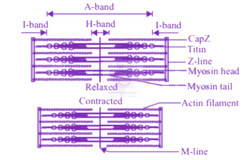
Related Theory Myofibrils are composed of long myofilaments of actin, myosin, and other associated proteins. These proteins are organized into regions termed sarcomeres, the functional contractile region of the myocyte. Within the sarcomere actin and myosin, myofilaments are interlaced with each other and slide over each other via the sliding filament model of contraction. The regular organization of these sarcomeres gives skeletal and cardiac muscle their distinctive striated appearance.